[ad_1]
Bone tissue labeling is not merely about learning and memorizing anatomical terms. It is about exploration of the dynamic, self-renewing tissue. In both clinical and academic settings, confusion between macroscopic layers and microscopic systems can lead to errors in clinical application and identification. A single orthopedic CT scan may contain hundreds of slices, while a whole-slide bone histopathology image can exceed several gigapixels. Accurately identifying cortical bone, trabecular bone, marrow spaces, fractures, osteoid tissue, implants, and pathological lesions requires deep anatomical understanding and rigorous quality control.
To gain a better understanding of annotating skeletal biology, keep reading this blog. The guide explains the complete bone annotation process, key challenges, quality standards, and the tools that enable scalable medical AI development.
Why Bone Tissue Annotation is Demanding for Medical AI
Bone tissue annotation is not merely a data preparation step. It directly influences clinical reliability, regulatory readiness, and model accuracy. AI systems trained on well-annotated bone datasets can support:
- Fracture detection and classification
- Osteoporosis screening and bone density assessment
- 3D surgical planning and navigation
- Bone tumor detection and segmentation
- Joint degeneration analysis
- Implant positioning and post-operative assessment
- Digital pathology for osteosarcoma and marrow disorders
Understanding Bone Tissue Structures
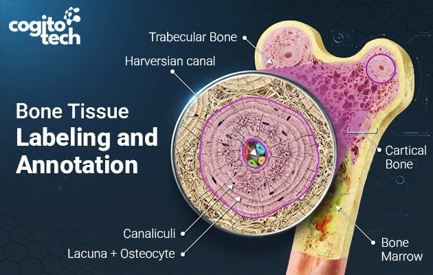
Broadly, bone tissue is categorized into two types: compact and spongy bone. Compact bone, also referred to as cortical bone, comprises the dense and hard outer layer of a bone, providing protection and strength. Spongy bone, also known as trabecular or cancellous bone, is less dense and lighter than compact bone. It exists in the interior of bones, at the ends of long bones, within flat and vertebral bones.
How Key Bone Structures are Annotated
Before annotation starts, a clear labeling ontology is crucial.
Cortical Bone
The cortical bone is the dense outer shell. It provides structural strength and is often the primary focus in fracture analysis.
Example – In a CT scan of the femur, annotators use semantic segmentation (2D or 3D voxel masks) to outline the cortical boundary across all slices.
Reason to choose semantic segmentation – It is chosen because the cortical layer forms a continuous anatomical structure. Pixel- or voxel-level labeling enables AI models to measure cortical thickness, discover areas of cortical erosion, detect subtle fractures, and perform bone assessment (which is not possible with simple bounding boxes).
Trabecular Bone
The trabecular bone is the porous inner network that is extremely relevant for osteoporosis and metabolic bone disease assessment.
Example – In high-resolution CT or micro-CT imaging, the trabecular region is labeled separately from cortical bone.
Reason to choose 3D volumetric segmentation – Trabecular bone has a complex 3D lattice structure. 3D volumetric segmentation is a preferred tool as it preserves the spatial arrangement of trabeculae, allowing AI models to evaluate bone mineral density, connectivity, trabecular spacing, and structural integrity.
Bone Marrow
Bone marrow is the soft tissue within the medullary cavity. It is commonly evaluated in MRI and pathology studies.
Example – In MRI scans of the spine, annotators label regions showing marrow edema or infiltration.
Reason to choose Region segmentation (MRI) or polygon annotation (pathology) – Marrow abnormalities appear as regional signal changes rather than sharply defined objects. Region-based segmentation is preferred because it captures the extent and distribution of abnormal marrow signal, which is more clinically meaningful than a tight object boundary.
Osteoid
Osteoid is newly formed, unmineralized bone matrix. It is particularly important in pathology and tumor evaluation.
Example – In a whole-slide histopathology image of osteosarcoma, pathologists draw polygon annotations around osteoid-producing tumor regions.
Reason to choose polygon annotation on whole-slide images – Osteoid areas often have irregular microscopic boundaries, requiring expert interpretation. Polygon annotation offers the flexibility recommended to precisely follow these complex contours. It helps AI models to distinguish malignant osteoid from normal bone and surrounding stromal tissue.
Fracture Lines
Fracture lines are discontinuities in bone that may be complete, incomplete, displaced, or occult.
Example – In an X-ray of the distal radius, annotators may place a bounding box around the fracture or draw a polyline along the fracture line.
Reason to choose bounding for detection; polyline/polygon for precise fracture mapping
- Bounding boxes are used when the goal is rapid fracture detection and training object-detection models.
- Polylines or polygons are chosen when precise localization is required for fracture classification, displacement measurement, or surgical planning.
Bone Lesions
Bone lesions are abnormal areas of bone tissue that can be malignant or benign. They are caused by infection, trauma, metabolic disorders, or tumors.
Example – In a CT scan of a vertebral body, annotators outline the lesion using instance segmentation.
Reason to choose instance segmentation or 3D lesion segmentation – Bone lesions are treated as individual pathological entities that must be separated from surrounding normal bone and from other lesions. Instance segmentation enables AI models to calculate lesion volume, shape, margins, and progression over time, which is essential for oncology and treatment-response assessment.
Technical Challenges Faced in Bone Tissue Labeling
Bone tissue annotation is more complex than generic images due to the combination of fine anatomical structures, disease-related changes, imaging artifacts, and the sheer scale of 3D medical datasets. The following challenges affect annotation accuracy, which further impacts the reliability of AI models.
Thin Cortical Boundaries
The cortical bone appears as a thin and high-density rim, particularly in pediatric scans, small bones, or low-dose CT studies. In some slices, the cortex may be only 2-4 pixels thick, making it difficult to determine the exact boundary between cortical bone and surrounding soft tissue.
A slight error in boundary placement can affect:
- cortical thickness measurements,
- bone mineral density calculations,
- fracture detection sensitivity,
- And surgical planning models.
Due to these impacts, rather than relying on a single slice, annotators have to review the structure across multiple coronal, axial, and sagittal slices. It increases annotation time and requires substantial anatomical expertise.
Metal Artifacts from Orthopedic Implants
Orthopedic implants such as plates, screws, rods, and joint prostheses can produce streak artifacts and beam-hardening effects in CT images. These artifacts create bright and dark bands that may obscure adjacent bone structures, fracture lines, or areas of bone healing.
For annotators, this means:
- the true bone boundary may not be visible,
- fracture margins may be partially hidden,
- and automated segmentation tools may incorrectly include artifact regions as bone.
As a result, implant cases often require manual slice-by-slice correction and close review by experienced radiologists or orthopedic specialists. These cases are also important for AI training because models deployed in clinical practice must learn to operate reliably in the presence of hardware.
Osteoporotic and Low-Density Bone
In osteoporotic patients, the cortical bone becomes thinner and less dense, while the trabecular network loses its normal structure. The contrast between cortical and trabecular bone is therefore much less distinct than in healthy bone.
This creates two major annotation challenges:
- Boundary ambiguity – it becomes arduous to determine where the cortex ends and trabecular bone begins;
- Inconsistent labeling – different annotators may choose different thresholds or anatomical interpretations.
These ambiguities can introduce bias into the dataset and reduce the accuracy of AI models designed for osteoporosis assessment or fracture risk prediction.
Inter-Observer Variability
Bone tissue annotation is not always objective. Two experienced radiologists may interpret the same fracture margin, lytic lesion, or callus boundary differently, especially when the abnormality is subtle.
For instance:-
one expert may include surrounding bone edema in the annotation,
while another may label only the visible fracture line.
This variability affects:-
- dataset consistency,
- model training stability,
- and the reproducibility of research results.
To minimize this issue, high-quality annotation projects use:
- standardized annotation protocols,
- consensus reviews,
- adjudication workflows,
- and agreement metrics such as Dice Similarity Coefficient (DSC) and Cohen’s kappa.
Large 3D Imaging Volumes
Modern CT studies can contain from 64 to 640+ slices, and each slice may require detailed review. A single pelvic or spinal case can take several hours to annotate accurately if performed entirely manually. The challenge extends beyond the number of slices, as it demands maintaining spatial consistency across the entire 3D volume. A boundary that is shifted by a few pixels in consecutive slices can create unrealistic 3D geometry and negatively affect downstream applications such as:
- surgical navigation,
- implant design,
- biomechanical simulation,
- and 3D printing.
Annotation teams often combine the following to make large-scale projects successful:-
- semi-automatic segmentation,
- AI-assisted pre-labeling,
- batch quality checks,
- and human-in-the-loop validation workflows.
Bone Structure, Annotation Type, and Rationale
| Structure | Annotation Type | Rationale |
|---|---|---|
| Cortical bone | Semantic segmentation | Continuous boundary and quantitative thickness analysis |
| Trabecular bone | 3D volumetric segmentation | Preserves complex 3D architecture |
| Bone marrow | Region segmentation | Captures diffuse signal abnormalities |
| Osteoid | Polygon annotation | Follows irregular microscopic contours |
| Fracture lines | Bounding box/polyline | Balances detection speed and localization precision |
| Bone lesions | Instance segmentation | Separates individual lesions for volumetric analysis |
Tools Commonly Used for Bone Annotation
Bone tissue labeling projects involve different tools rather than a single platform. The choice depends on imaging modality (MRI, CT, X-ray, or histopathology), required precision level, and whether the project is intended for clinical AI development or research.
3D Slicer – Advanced Volumetric Bone Segmentation
3D Slicer is a widely used open-source platform for orthopedic imaging and 3D medical image analysis. It is mostly preferred for CT-and MRI-based bone segmentation because it supports native DICOM workflows, multi-planar visualization, and voxel-level editing.

Why it is used for bone annotation
- Hounsfield Unit (HU) thresholding allows rapid isolation of cortical bone from surrounding soft tissue.
- 3D volumetric segmentation preserves the spatial relationship between bones, joints, and fracture fragments.
- Multi-planar review (axial, coronal, sagittal) helps annotators maintain boundary consistency across the entire scan.
- Built-in surface reconstruction and mesh generation support surgical planning and biomechanical analysis.
- Python scripting enables workflow automation and integration with MONAI-based AI pipelines.
Best suited for femur, pelvis, spine segmentation, complex fracture reconstruction, and pre-operative planning.
ITK-SNAP – High-Precision Manual Refinement
ITK-SNAP is designed for precise manual and semi-automatic segmentation. While it is lighter than 3D Slicer, it is often used when experts need fine control over difficult boundaries.

Why it is used for bone annotation
- Active contour (snake) segmentation can follow complex bone edges with minimal manual input.
- Voxel-level editing tools allow correction of thin cortical boundaries that automated algorithms might miss.
- The interface is optimized for rapid slice-by-slice refinement. This makes it useful for challenging fracture cases.
Best suited for fracture boundary correction, osteoporotic bone segmentation, tumor margin refinement, and small research studies requiring expert-reviewed annotations.
QuPath – Digital Pathology and Osteoid Annotation
QuPath is a specialized platform for whole-slide pathology image analysis. It is essential when bone annotation moves from radiology to microscopic tissue evaluation.

Why it is used for bone annotation
- Supports gigapixel whole-slide images without requiring image downsampling.
- Polygon and brush tools allow accurate outlining of irregular osteoid and tumor regions.
- Built-in cell detection and tissue classification help quantify marrow composition, fibrosis, and necrosis.
- Scripting capabilities enable batch analysis and reproducible pathology workflows.
Best suited for osteoid annotation, bone marrow analysis, tissue classification, and quantification.
MONAI Label – AI-Assisted Bone Annotation
MONAI Label is an AI-assisted annotation framework that integrates with tools such as 3D Slicer. Instead of annotating from scratch, experts review and correct model-generated segmentations.

Why it is used for bone annotation
- AI generates an initial segmentation mask, minimizing manual effort.
- Expert corrections are fed back into the model, enabling continuous learning and improvement.
- Supports human-in-the-loop workflows, which are critical for regulatory-grade medical datasets.
- Reduces annotation time for large 3D orthopedic studies.
Best suited for large-scale orthopedic datasets, multi-site annotation projects, and product-grade human-in-the-loop validation.
How Cogito Tech Ensures Bone Tissue Annotation Quality
Medical AI datasets demand a higher level of precision than generic computer vision datasets. A few pixels of error in a cortical boundary or fracture margin can affect lesion volumes, bone measurements, and even surgical planning. To ensure high annotation accuracy and reproducibility, Cogito Tech follows a multi-layer quality management framework designed specifically for orthopedic imaging and digital pathology projects.
Multi-Layer Quality Assurance Workflow
- Primary annotation is performed by trained medical annotators with expertise in musculoskeletal imaging and pathology workflows.
- Secondary QA review checks boundary accuracy, label completeness, slice-to-slice consistency, and adherence to project guidelines.
- Expert validation is conducted by radiologists or pathologists for complex fractures, tumors, osteoid regions, and ambiguous findings.
- A final audit verifies dataset integrity, annotation formats, metadata consistency, and export readiness for AI training pipelines.
Measuring Annotation Consistency
- Dice Similarity Coefficient (DSC) measures overlap between segmentation masks.
- Intersection over Union (IoU) evaluates boundary agreement and spatial accuracy.
- Cohen’s kappa assesses agreement for categorical labels such as fracture type or lesion classification.
Standardized Annotation Protocols
To minimize interpretation differences, Cogito Tech develops project-specific standard operating procedures (SOPs) that define:
- cortical and trabecular boundary rules,
- fracture inclusion and exclusion criteria,
- minimum lesion size thresholds,
- implant and hardware labeling conventions,
- pathology tissue hierarchies,
- and handling of ambiguous or artifact-affected regions.
Preserving Metadata and Traceability
Annotation quality is not limited to the masks themselves. All DICOM metadata, including scanner parameters, voxel spacing, orientation, and study identifiers. It is preserved throughout the workflow. This supports:
- accurate downstream modeling,
- reproducibility of research results,
- audit trails,
- dataset version control,
- and regulatory documentation for FDA, MDR, and other compliance frameworks.
Why Healthcare AI Teams Choose Cogito Tech
Cogito Tech provides end-to-end medical data annotation services for radiology, orthopedics, and digital pathology.
Our capabilities include
- DICOM, NIfTI, and whole-slide image annotation
- 2D and 3D bone segmentation
- Fracture and implant labeling
- Bone tumor and lesion annotation
- Pathologist-guided histopathology labeling
- Human-in-the-loop validation
- HIPAA- and GDPR-aligned workflows
Conclusion
The success of bone tissue labeling and annotation depends on far more than annotation software. It requires anatomical expertise, modality-specific workflows, standardized ontologies, and rigorous quality assurance. Whether you are building AI for fracture detection, osteoporosis assessment, surgical planning, bone tumor analysis, or digital pathology, investing in a structured annotation pipeline is essential for achieving clinically reliable and scalable AI performance.
At Cogito Tech, we help healthcare AI teams transform complex bone imaging data into high-quality, production-ready training datasets.
Frequently Asked Questions (FAQ)
Bone tissue labeling is the annotation of anatomical structures such as bone marrow, bone lesions, fractures, osteoid tissue, cortical bone, and trabecular bone. The main goal of bone tissue labeling is to train and validate artificial intelligence (AI) models for orthopedic imaging and digital pathology.
CT, MRI, X-ray, and whole-slide histopathology images can be used for bone tissue annotation. For marrow and soft-tissue analysis, MRI is preferable; for pathology slides, for tumor and osteoid evaluation; for X-ray, for fracture recognition and 3D bone segmentation.
The challenge in bone annotation is represented by thin cortical boundaries, osteoporotic bone with poor contrast, metal artifacts from implants, the complexity of 3D anatomy, and large volumes of imaging. All these elements need medical expertise and multi-stage quality assurance.
Consistency is maintained by standard annotation guidelines, calibration sessions, a double-review workflow, expert adjudication, and consistency metrics like Dice Score, IoU, and Cohen’s kappa.
Bounding boxes help track fractures quickly, but they do not reflect the exact fracture shape. Polygons or polylines are chosen for fracture localization, classification, displacement measurement, and surgical planning.
DICOM metadata includes voxel spacing, image orientation, scanner parameters, study identifiers, etc. Preservation of DICOM metadata is critical for proper measurements, reproducible research, regulatory documentation, and AI training pipelines.
[ad_2]
Source_link