Medical imaging AI is transforming healthcare. Behind every diagnostic model that detects lung nodules and brain hemorrhages, flags a fracture, or segments an organ lies thousands of carefully annotated medical images. And the vast majority of those images are in one format: DICOM.
DICOM image annotation is not a task that generic image labeling workflows can easily accommodate. It is far more than drawing boxes around abnormalities. DICOM medical image annotation demands clinical expertise, an understanding of complex imaging metadata, specialized tools, and a fundamentally different approach to quality — because in medicine, annotation errors don’t just affect model performance. They put patients at risk.
This article explores every aspect of DICOM medical image annotation, including how it differs from conventional image labeling, common use cases, challenges, annotation workflows, tools, and how specialized medical AI innovation hubs accelerate development.
DICOM stands for Digital Imaging and Communications in Medicine. It is the global standard for storing, transmitting, and managing medical images and the metadata associated with them.
DICOM is recognized as the ISO 12052 standard and supports every major imaging modality — X-ray, CT, MRI, ultrasound, PET, and mammography. DICOM has expanded far beyond radiology and is widely used across ophthalmology, dentistry, cardiology, and other medical specialties. There are now billions of DICOM images in clinical use worldwide, flowing through hospitals, imaging centers, and research institutions, and healthcare networks every day.
Unlike standard image files, each DICOM files are metadata-rich by design. They embed patient identifiers, scanner acquisition parameters, study and series information, and clinical workflow information alongside the pixel data. This enables interoperability — a DICOM image acquired on one manufacturer’s scanner can be reviewed and interpreted using any DICOM-compliant system anywhere in the world. At the same time, DICOM annotation is considerably more complex than any other type of image labeling.
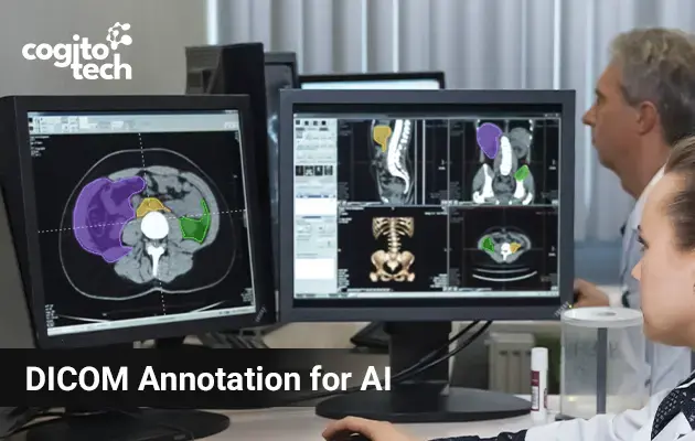
DICOM annotation is the process of adding labels and markings to DICOM medical images to highlight specific regions of interest — tumors, fractures, anatomical structures, lesions, organ boundaries — for clinical AI training and validation.
Experts mark where the nodule begins and ends, classify it as malignant or benign, and record its dimensions to turn raw pixels into actionable insight. Annotated images become training data that enables AI systems to detect similar findings at clinical speed and scale.
Annotation is essential for multiple healthcare and AI workflows: AI model training, diagnostic validation, surgical planning, clinical research, and regulatory submission. The same labeled dataset can help meet FDA evidentiaryrequirements while also informing a surgeon’s pre-operative planning.
How DICOM Annotation Differs from Standard Medical Image Annotation
Not all medical images are DICOM. Healthcare AI workflows also rely on formats such as JPEG, PNG, NIfTI, and Whole Slide Imaging (WSI). Understanding where DICOM sits relative to these formats is essential because DICOM studies introduce an entirely different level of complexity, making their annotation a distinct discipline.
| Aspect | DICOM Annotation | Standard Image Annotation |
|---|---|---|
| Data Structure | Multi-layered medical studies | Single images |
| Dimensions | Often 3D or 4D volumes | Primarily 2D |
| Metadata | Extensive clinical metadata | Minimal metadata |
| Measurements | Clinically calibrated | Pixel-based |
| File Relationships | Study-Series-Instance hierarchy | Independent files |
| Required Expertise | Clinicians and radiologists | General annotators often sufficient |
| Compliance Requirements | HIPAA, GDPR, PHI management | Lower complexity |
| Workflow Integration | PACS and hospital systems | Standalone workflows |
For example, a CT scan may contain hundreds or even thousands of slices. Marking a single lesion may require
reviewing an entire 3D volume rather than a single image.
Annotation Types Used in DICOM Imaging
Annotation tasks vary based on clinical tasks. The most common types in DICOM annotation workflows include:
Bounding Boxes: Annotators draw rectangular bounding boxes around anatomical structures or abnormalities. Used to localize findings like tumors or fractures for detection models.
Polygons: Used to trace the exact contours of irregular anatomical structures such as tumor margins, organ boundaries, and unusually shaped lesions. More time-consuming than bounding boxes but significantly more anatomically reliable.
Semantic Segmentation: Pixel-level labeling that assigns a class to every pixel in the image, used for organ segmentation, tissue classification, and lesion delineation.
3D Volumetric Segmentation: Assigning voxel-level labels for organs, tumors, and structures through all planes. This is the most used and clinically accurate form of DICOM annotation.
Classification Labels: Tagging images or regions (e.g., normal, malignant, and benign) to enable supervised classification models.
DICOM Annotation Use Cases
Radiology and Disease Detection
Annotated DICOM images are extensively used in radiology and disease detection to characterize abnormalities across every imaging modality, such as identifying fracture locations and severity on X-rays, highlighting tumor boundaries in CT and MRI images, and analyzing heart function and vascular abnormalities in echocardiography and cardiac MRI.
Oncology and Tumor Analysis
Annotated DICOM images support the entire cancer AI pipeline – from detection to treatment monitoring. For example, annotated mammography data enables AI models to detect early-stage malignancies. While segmentation models can assess tumor size and track response to chemotherapy. Multi-modal annotation (e.g., the combination of PET and CT studies) enables AI systems to analyze both the structure and metabolic activity of tumors simultaneously, providing a comprehensive picture for staging and treatment planning.
Cardiology
Annotated DICOM data, including echocardiograms, cardiac CT, and cardiac MRI, is used to train cardiac imaging AI. Tasks such as chamber segmentation, measurement of ejection fraction, coronary artery stenosis identification, and detection of structural abnormalities are done on DICOM images. They reduce inter-observer variability between cardiologists and speed up time-sensitive assessments.
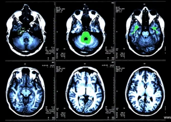
Neurology and Brain Imaging
Annotated brain MRI scans support AI systems for lesion detection in multiple sclerosis, tumor segmentation for surgical planning, and stroke infarct volume measurement. The complexity of neuroanatomy connects annotator expertise to data quality.
Surgical Planning
Annotated DICOM data from CT and MRI scans can be used to generate detailed 3D anatomical models – highlighting tumors, healthy muscles, and organs with clinically accurate precision for pre-operative planning. These 3D reconstructions allow clinicians to rehearse the entire operation before entering the operating room.
DICOM Image Annotation: Process and Challenges
DICOM data annotation is different from standard computer vision data labeling – more complex, time-intensive,
and expertise-driven.
The annotation process includes the following steps:
- Data preparation and de-identification: Before annotation, protected health information (PHI) must be removed from DICOM files while preserving the clinically relevant metadata. Patient names, Social Security numbers (SSNs), dates of birth, health record numbers, and other identifiers can appear in headers, in burned-in pixel data overlays, and in reconstructable structures.
- Protocol definition: This involves precisely defining what structures or abnormalities are to be annotated, specifying annotation methods, such as segmentation, bounding box, or landmark, and documenting ground-truth examples. Moreover, edge cases, ambiguous findings, and variability resolution procedures must be communicated in writing before annotators start working.
- Visualization and windowing: DICOM images contain far more information than is visible on a standard screen. Windowing is used to adjust the contrast and brightness mapping of the pixel data to optimize the visibility of structures relevant to a given annotation task. Proper windowing is essential for accurate annotation, a skill that general-purpose annotators don’t have.
- Slice-by-slice annotation: Annotators use multiplanar reconstruction (MPR) to work through each relevant slice, marking regions of interest that must remain consistent across the volume, to achieve spatial accuracy across all three dimensions.
- Quality review: DICOM datasets are reviewed in multi-tiered cycles – annotator, senior reviewer, radiologist. Inter-annotator agreement metrics are tracked to discover systematic inconsistencies.
Challenge
- Volume and Complexity: A single CT or MRI study can contain hundreds of slices. Slice-by-slice annotation is data-intensive and time-consuming. Volumetric segmentation may require an annotator to trace findings consistently across dozens or even hundreds of slices. Region of interest, such as tumors, fractures, anatomical structures, and lesions, may appear in only a small subset of slices within a large study, demanding close attention throughout the process.
- Expert Dependency: Unlike standard computer vision projects, DICOM medical image annotation requires clinical interpretation. For radiologists, finding time for annotation amid demanding clinical workloads can be challenging.
- Inter-Observer Variability: DICOM image interpretation often involves a degree of subjectivity. Even two experienced radiologists may delineate lesion boundaries slightly differently or may not agree on subtle findings. Implementing consensus-based workflows, annotation guidelines, and multi-stage review processes is essential to reduce variability.
- Regulatory Compliance: DICOM files may contain protected health information, including burned-in pixel overlays, which must be removed before datasets can be used for model training to comply with HIPAA, GDPR, and other data governance frameworks. Protected information can appear in unexpected locations within a DICOM file, and incomplete de-identification may expose patient data. Data vendors must maintain secure and auditable workflows.
- Scalability: Due to the volumetric nature of the data and clinical complexity, maintaining annotation quality across thousands of DICOM studies becomes increasingly difficult. Scaling annotation operations requires not only domain expertise but also standardized workflows, robust quality assurance processes, and efficient project management.
How Cogito Tech’s Medical AI Innovation Hub Supports DICOM Annotation
Cogito Tech’s Medical AI Innovation Hubs are designed for clinical expertise, operational infrastructure, and regulatory discipline. The Hubs combine a multidisciplinary team of board-certified radiologists, CCTA readers, and other medical professionals drawn from hospital networks worldwide. Domain experts benchmark and validate annotation across specialties with the breadth of perspectives that reduces bias and improves label accuracy across diverse patient populations and imaging contexts.
Operating within HIPAA-, GDPR-, and ISO 27001-aligned environments, the Hubs support the creation of audit- ready medical imaging datasets while maintaining strict controls around patient privacy and data governance. These capabilities align particularly well with DICOM annotation projects involving tumor segmentation, organ segmentation, clinical validation, PHI/PII removal, human-in-the-loop quality review, and AI model evaluation and refinement.
DataSum, our proprietary data transparency framework, brings transparency to medical AI training data – providing structured, auditable insight into the composition, quality, and coverage of labeled datasets. Combined with HIPAA- compliant, FDA-ready, and 21 CFR Part 11-aligned workflows, this gives medical AI teams the documentation infrastructure they need to support regulatory submissions, not just model training.
Conclusion
DICOM medical image annotation forms the foundation of modern healthcare AI. Unlike standard image labeling, it requires specialized expertise, stricter regulatory compliance, and the ability to manage large, complex imaging datasets. Inaccuracies and inconsistencies do not simply degrade model performance; they can contribute to diagnostic errors at scale.
However, with the right combination of clinical expertise, rigorous quality assurance, and purpose-built tooling, DICOM annotation enables the development of transformative medical AI systems that can detect cancer earlier, triage patients more accurately, and support clinicians in making faster, more informed decisions.