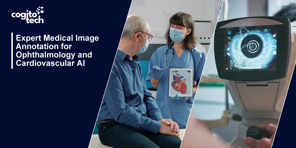
Fields like ophthalmology and cardiovascular care, where imaging is central to diagnosis, have emerged as critical frontiers for AI, making high-quality medical image annotation services indispensable. It’s about training medical AI systems with data that shows how doctors actually think and make decisions. This means the data needs to be labeled by experts, such as board-certified ophthalmologists, retinal specialists, and cardiovascular imaging experts, who really understand the importance of each label they assign.
Cogito Tech offers high-quality medical image annotation services specifically designed with the unique requirements of two of the most precise fields of medicine. Through this blog, we will explore how our medical image annotation services are delivered by a team of board-certified clinicians, using modality-specific workflows and an added layer of quality assurance.
Decoding Retinal Complexity in Ophthalmology AI
Ophthalmology is one of the highest-stakes domains in medical AI. The eye is a small organ with subtle pathological features, and the margin for labeling error is essentially zero. If you’re building a diagnostic model for retinal disease, the quality of your labeled dataset isn’t a background variable — it’s the primary determinant of clinical validity.
What precise annotation actually involves:
Image Classification
Labeling helps AI models identify and distinguish eye conditions, such as diabetic retinopathy, macular degeneration, and glaucoma. This allows ophthalmologists to assess and prioritize cases in a timely and appropriate manner.
Object Detection
Object detection in ophthalmology involves labeling objects or features within an image, such as blood vessels or optic nerve heads. Precisely labeled datasets enable AI models to locate and identify these objects, helping detect abnormalities or pathologies. For example, detecting and measuring blood vessel diameters can aid early diagnosis of hypertensive and diabetic retinopathy.
Segmentation
Segmentation involves labeling specific regions or structures within an image, such as the retina or optic nerve. Precisely labeled datasets train AI models to segment these regions accurately, enabling detailed analysis. For example, segmenting the retina helps quantify retinal layer thickness and identify thinning or thickening that may indicate disease progression.
Optical Coherence Tomography (OCT) Labeling
OCT labeling focuses on segmenting and classifying features within OCT images, such as retinal layers, the optic nerve head, or the choroid. Precisely labeled datasets train AI models to identify structures and anomalies in OCT scans. For example, labeling retinal layers helps detect thickness changes and identify layers affected by conditions like macular edema or retinal detachment.
Fundus Image Labeling
Fundus image labeling involves annotating features like the optic disc and blood vessels. Precisely labeled datasets enable AI models to identify and analyze these structures. For example, labeling the optic disc helps measure its size, detect abnormalities such as optic disc drusen or swelling, and monitor changes over time.
Precision Annotation for Cardiovascular AI Models
ECG (Electrocardiogram) Labeling
Expert annotators help to identify cardiac conditions such as arrhythmias, heart block, and transient episodes of myocardial ischemia. They are skilled at accurately identifying all abnormalities on the ECG and labeling them, including any irregular heart rhythm, new ST-segment changes, and abnormalities of the P-QRS-T waveforms. ECG annotations enable healthcare professionals to have a more complete picture of an ECG when making diagnoses, monitoring, and treating patients with known and unknown heart conditions.
Cardiac CT Labeling
We use X-rays to visualize the heart and coronary arteries using cardiac CT. We provide annotated cardiac CT datasets derived from clinically acquired scans. The coronary arteries are accurately segmented and classified into specific segments. Any detected stenoses, calcifications, etc., are annotated and described. Other structures that are annotated and accurately labeled are the ventricles and atria, the heart valves (aortic and mitral), the cardiac connections, and other relevant cardiac structures. This allows assessment and visualization of coronary artery disease, as well as evaluation of cardiac morphology to determine whether any abnormalities are present. Accurate segmentation and labeling allow for accurate cardiac diagnosis and treatment planning.
Wearable Sensor Labeling
With the growing popularity of wearable technology, there is a growing need for high-quality annotated data from sources such as smartwatches and fitness trackers. It is not enough for annotators to simply label heart rate for rest and exercise states; annotators label activity states and signal segments, enabling downstream computation of metrics like heart rate variability (HRV). This data is invaluable to health practitioners and individuals alike and can be used to monitor cardiovascular health and track wearer activity levels, ensuring they are doing everything in their power to live healthily.
Echocardiography Labeling
Echocardiographic images show heart structures in motion, and doctors use them to evaluate heart anatomy and function. Our large number of trained annotators can precisely segment and annotate the heart chambers (left and right ventricles, left and right atria), heart valves (mitral and aortic), and vessels (aorta and pulmonary artery). Accurate annotations of echocardiographic images facilitate cardiac evaluation and help doctors and clinicians make accurate diagnoses and treatment decisions.
Why Choose Cogito Tech?
More than annotation, if you are looking for a clinical data infrastructure partner, Cogito Tech is your answer. Most annotation vendors hand you labeled files and walk away. Cogito Tech is built differently. From the moment your imaging data enters our platform to the final validated dataset delivered to your ML team, every step is designed around one outcome: training data that makes your medical AI deployable, regulatory survival, and clinically reliable.
Clinical credibility
All our imaging data, retinal fundus, OCT, echocardiograms, and coronary CTs are managed by our expert team, who annotate and are verified by licensed clinicians (doctors, nurses, and specialists); they are not crowdsourced. Our datasets integrate seamlessly without disrupting your existing pipeline. We support DICOM-compliant proprietary formats with robust version control, ensuring the credibility of our compliant-ready datasets.
Risk reduction
There are different risk types that undermine the model’s credibility, including patient safety risks when wrong predictions harm patients, regulatory risks when data is not audited, legal risks that could lead to liability for adverse outcomes, and commercial risks when hospitals do not adopt the model, which could lead to loss of trust and sales. Non-credible data (scraped, imbalanced, unvalidated) often carries the above risks, but clinically credible data means you can prove the model was built on a sound foundation.
Global workforce domain-experts
Across geographically distributed teams, we work with data scientists, medical experts, and digital leads through a cloud-based environment built for healthcare data compliance. Everyone works from the same source of truth, with role-based access controls and audit trails intact. Board-certified radiologists, CCTA readers, and ophthalmic graders are embedded into the annotation workflow from the start— not an afterthought.
Production scaling
We offer data that are recent enough to remain relevant as the model scales into production over months and years, and are structured and validated specifically to support enterprise-grade production environments. Clients who have used our datasets in production report consistent model performance even as volume and complexity increased.
Regulatory survival
Annotated datasets are delivered in the format required by our client’s ML pipeline and with the documentation your regulatory submission demands. Cogito Tech provides not only labeled data but also organized training sets that can be checked and approved for clinical use, and 21 CFR Part 11 (audit trails) is also crucial. As per the client’s needs, we offer JSON, XML, DICOM-SR, COCO, Pascal VOC, custom format, FDA SaMD-aligned, and clinical validation-ready.
At Cogito Tech, our AI data solutions have built clients’ systems to deliver clinically, operationally, and regulatorily viable results. If you’re building medical AI that needs to perform in real clinical environments, the training data infrastructure matters as much as the model architecture.
Conclusion
The difference between how well a medical AI performs in demo mode and in real-world practice, where patient lives are at stake, is what AI models use as training datasets when running algorithms. Between high-quality and low-quality training data, a model must undergo multiple runs and tests.
In ophthalmology and cardiovascular fields, as we discussed in this blog, errors in the boundaries of a structure or in the classification of a wall motion segment can have serious clinical implications. There is no room for error in life-critical specialties, and keeping the annotation insightful is crucial for proper model training. Cogito Tech works to provide a solid data foundation, as medical AI needs reliable, compliant, and high-quality training data. Reach out to discover how we can help.